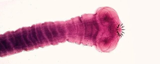
|
Stem cells are transforming medical research, promising a clinical revolution in which doctors will employ embryonic and adult stem cells to repair failing hearts, create new organs and tissues, restore brains damaged by stroke or neurodegenerative disorders, and treat hereditary defects.
However, before stem cells can realise their promise, researchers must develop reliable systems to maintain and multiply them in culture and learn how to manipulate the microenvironment to direct cells into selected developmental pathways.
In other words, they must create particular microenvironments which imitate the real environments in which they would function in the body and in which stem cell research can be conducted.
To this end, Professor Min Gu and Dr Daniel Day, of the Centre for Micro-Photonics at Swinburne University of Technology, are developing microfluidic ‘lab-on-a-chip’ devices.
Little larger than a microscope slide, the microfluidic chips will contain tiny bioreactors, fed by microfluidic ‘plumbing’ that will supply controlled quantities of nutrients and cell growth factors from on-board reservoirs, mimicking the natural milieu in which stem cells replicate and differentiate into other cell types in the body’s tissues and organs.
Professor Gu says the chips are being designed to culture embryonic stem cells, which can differentiate into any of the 210-odd different cell types in the body. They will also be used to replicate adult haemopoietic stem cells, which give rise to the specialised cells of the blood and immune systems.
The Swinburne component is part of a major development program for stem cell research involving the Australian Stem Cell Centre at Monash University, the Monash University Centre for Green Chemistry, CSIRO Molecular and Health Technologies, and the Cooperative Research Centre for Polymers.
Professor Gu says the aim is to create designs for microfluidic chips that can be mass-fabricated at low cost. Programmable and almost maintenance-free, they will allow researchers to shrink macroscopic laboratory experiments to Lilliputian dimensions.
Arrays of multi-chambered microfluidic chips will allow multiple experiments to be run in parallel. Researchers will be able to experimentally adjust each biochemical or physical parameter in the bioreactors, and observe how stem cells respond to different concentrations and combinations of nutrients and cytokines, as well as changes in temperature, pressure or oxygen levels.
Professor Gu says the chips will help researchers determine the conditions required to maintain embryonic and adult stem cells in an undifferentiated state, and how to control their differentiation into other cell types.
Associate Professor David Haylock, a senior research scientist with the Australian Stem Cell Centre, says that if the project is successful, it will take stem cell research and its clinical applications to the next stage.
"There’s no good technology for growing stem cells and their progeny at the scale required for research or medical use," Dr Haylock says. "The bioreactor would allow us to grow stem cells and explore their full potential."
Microfluidic chips will allow researchers to conduct complex experiments under highly controlled conditions that would normally require costly, large-scale cell-culture equipment and monitoring devices.
Where other research groups and companies are developing microfluidic chips made of glass, silicon or polydimethylsiloxane using photolithography processes, the Swinburne researchers have developed a manufacturing process using femtosecond lasers (a femtosecond is a thousand-trillionths of a second) to fabricate the masters from which the chips can be produced in a variety of polymers.
The technique, called ‘two-photon ionisation’ focuses a high-energy femtosecond pulse laser into the target substrate material, which can be made from metal, glass or polymers. At the point where the laser beam is focused, the energy from the laser ionises the material, effectively removing it from the substrate. This method enables microscopic resolution features to be fabricated in the substrate, which can then be used as a master mould from which multiple copies can be replicated.
Computers programmed with digital templates for the chip’s components will steer the focus point of the laser pulses through the substrate, progressively building up complex three-dimensional shapes and cavities.
At lower energies, the focused beam can be used to polymerise a photosensitive resin, so structures can be etched into the chip surface, or constructed from the resin, creating complex 3D networks of microchannels and other microstructures.
Micropumps incorporated into the chips will pump precise quantities of nutrients or cell growth factors through microfluidic circuits into the bioreactor chambers containing the stem cells.
Professor Gu says the femtosecond lasers can also etch tiny optical gratings or 3D photonic crystals into the material that are extremely sensitive to optical changes in the cell microenvironment, such as changing temperatures, pH levels or other conditions in the bioreactor – including changes in the cells themselves.
By labelling antibodies with fluorescent molecules – fluorophores – that emit specific colours under ultraviolet light, researchers can observe the patterns of receptors expressed on the surface of the cells. The combinations of receptors will reveal whether the stem cells are in an undifferentiated state, or have begun to transform into other cell lineages – and, if the latter, what type of cell will emerge from the process.
Both the optical gratings and photonic crystals have been developed for another of the centre’s projects: to develop optical chips for telecommunication.
The centre has also developed laser-based techniques for manipulating micrometre-sized objects, which can be used to trap, observe and manipulate living cells as tiny as a red blood cell.
A tightly focused laser beam can trap cells at a focal point defined by the difference between the refractive index of the object and the surrounding medium. The immobilised cells can then be manipulated and studied.
Dr Haylock says thousands of the micro-scale bioreactors linked together would enable researchers to grow stem cells "with exquisite control" and in large numbers, representing a wide range of human genotypes.
"Ideally, for therapeutic applications, they will allow us to culture the recipient’s own cells," he says. This would avoid the risk of patients rejecting grafted cells, which happens if the donor and recipient have imperfectly matched immune-system genes – a problem that can affect organ transplants.
Dr Haylock says it is still not clear if embryonic stem (ES) cells will provoke rejection reactions or be universally suitable for patients. "ES cells are the great hope, and if there are rejection problems, perhaps we can match the immune-system type of the cell to the recipient."
He says the number of different ES cell lines in culture presently numbers in the "many 10s", so they represent only a small sample of the diversity of human genotypes. The bioreactor chip would be essential for maintaining a more comprehensive range of ES cell genotypes.
"Few of the existing ES cell lines are well-characterised biologically, and we are only able to grow a handful of specialised cell types from them.
"ES cells have the potential to differentiate into any of the more than 200 human cell types, but each will have different growth requirements. Microbioreactors will allow those conditions to be altered very precisely."
Dr Haylock says it is likely that rather than being grown in suspension, in solutions, the cells will be grown on micro-textured surfaces with nutrients and cell growth factors embedded in them.
The partners in the research consortium developing the microfluidic-chip bioreactors are already planning to establish a company to manufacture the chips.
At Swinburne’s Centre for Micro-Photonics, Dr Daniel Day explains: "We’re still in the pre-prototype phase, assessing a number of design aspects, but we should have a first-generation microfluidic chip some time next year."
He says the diverse, but complementary, skills offered by members of the consortium should create a range of commercial opportunities – for example, microfluidic chips with which drug developers can test experimental drugs on stem cells or differentiated cell lines.
A story provided by Swinburne Magazine. This article is under copyright; permission must be sought from Swinburne Magazine to reproduce it.
|